Radionuclide imaging
Creating radioactive tracers for PET and SPECT imaging will enable us to predict therapy effectiveness and outcomes as well as monitor response to radiotherapy. Tracers target the creation of new blood vessels (angiogenesis), macrophages and the biological response in terms of manganese concentrations. Equally, we are working on tracking of externally radiolabeled stem cells, macrophages, and T cells.
Example 1. Radiolabeled RGD peptides to image angiogenesis in the clinic
68Ga-labeled RGD peptide was first assessed in vitro for specificity to image angiogenesis in a preclinical tumour model (see Lobeek et al. 2018). The dimeric version of this tracer was then assessed in the clinic to visualise angiogenesis in head and neck cancer patients (Lobeek et al. 2020a, image on left) as well as patients with vascular malformations (Lobeek et al. 2020b).
Example 2. Radiolabeled RGD peptides to monitor angiogenesis response after radiotherapy
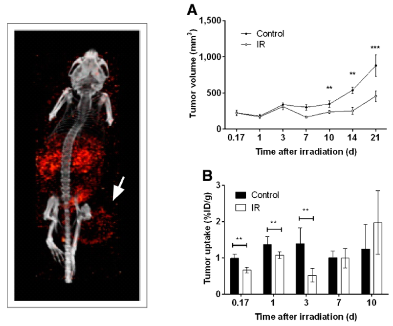
On the left we can see a SPECT image of an RGD peptide radiolabelled with In-111. The arrow is pointing to up uptake of In-111 in a tumour located in a mouse. This area is where new blood vessels have been formed, a process called angiogenesis. On the top right figure, we show that radiation can stop the rate at which tumours grow and that uptake of our tracer shows a response as early as 4 hours (0.17 days) after irradiation (bottom right). This suggests that radiolabelled RGD peptides can be used to predict tumour response to radiotherapy. From Terry et al 2015.

